Understanding Pleural Effusions
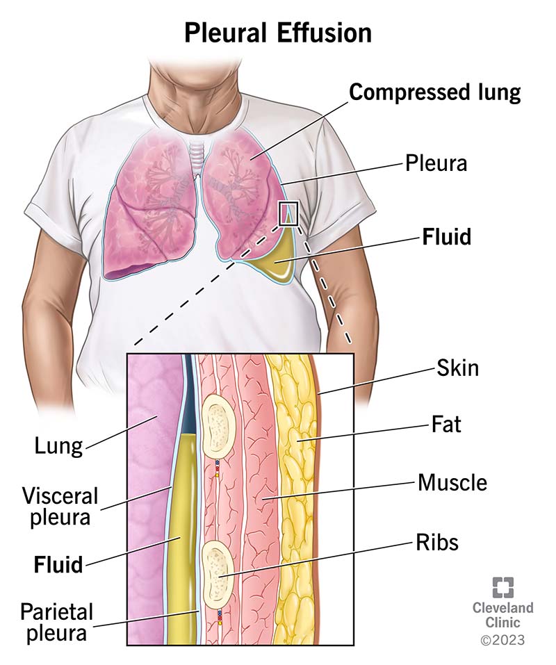
A pleural effusion occurs when excess fluid accumulates in the pleural space, the area between the lung tissue and the chest wall. Small amounts of fluid normally exist in this space to allow the lungs to move smoothly during breathing. However, when the balance between fluid production and reabsorption is disrupted, abnormal fluid accumulation can occur. Pleural effusions can develop from various underlying medical conditions and may range from asymptomatic to causing significant respiratory symptoms. Proper diagnosis and treatment of the underlying cause are essential for successful management and prevention of complications.
Pleural Disease Care at PulmoCrit
In-office ultrasound-guided thoracentesis — no hospital admission required for most diagnostic or therapeutic procedures
In-office chest ultrasound — real-time imaging to safely locate and characterize pleural fluid
Pleural fluid analysis — comprehensive laboratory coordination to identify infection, malignancy, or systemic cause
Recurrent effusion management — repeat thoracentesis or referral for indwelling catheter when appropriate
Same-week procedures — urgent cases expedited to minimize symptom burden and hospitalization risk
Available at Northridge, Encino, and Thousand Oaks. Request an appointment →
Causes of Pleural Effusions
Pleural effusions can develop from numerous underlying conditions, broadly categorized as transudative or exudative:
- Heart Failure: One of the most common causes of pleural effusions, particularly affecting the right side. As the heart's pumping ability decreases, fluid backs up into the lungs.
- Infection (Empyema): Bacterial, viral, or fungal infections of the pleura or lungs can lead to fluid accumulation. These are often associated with pneumonia.
- Malignancy: Cancer involving the pleura or lungs is a significant cause of pleural effusions. This represents advanced disease requiring specialized management.
- Pulmonary Embolism: Blood clots in the lungs can trigger pleural effusion development.
- Kidney Disease: Nephrotic syndrome and other renal conditions can lead to pleural effusions due to changes in fluid dynamics.
- Liver Cirrhosis: Advanced liver disease causes fluid retention and can result in pleural effusions.
- Connective Tissue Diseases: Conditions such as rheumatoid arthritis and lupus can cause pleural effusions.
- Pancreatitis: Inflammation of the pancreas can be associated with pleural fluid accumulation.
Symptoms and Diagnosis
Symptoms of pleural effusion vary depending on the size of the effusion and the underlying cause. Many small effusions are asymptomatic and discovered incidentally on imaging. Larger effusions typically cause shortness of breath, chest pain that worsens with breathing, dry cough, and reduced exercise tolerance. Diagnosis begins with clinical evaluation and imaging studies. Chest X-rays can detect moderate to large effusions, while ultrasound and CT imaging provide more detailed visualization. Pulmonary evaluation by our specialists includes assessment of your medical history and physical examination. To identify the underlying cause, thoracentesis (needle aspiration) is often performed to obtain pleural fluid samples for laboratory analysis.
Thoracentesis Procedure
Thoracentesis is a minimally invasive procedure in which a needle is inserted into the pleural space under ultrasound guidance to remove fluid samples for analysis. This procedure serves both diagnostic and therapeutic purposes. Diagnostically, the fluid is analyzed for characteristics such as protein content, cell counts, glucose, lactate dehydrogenase, and cultures to identify infections or malignancy. Therapeutically, removing excess fluid can provide immediate symptom relief and improve breathing. The procedure is generally safe and well-tolerated when performed by experienced pulmonary specialists, with minimal discomfort to the patient.
Treatment Options
Treatment of pleural effusions focuses on addressing the underlying cause while managing symptoms. Depending on the diagnosis, treatment may include:
- Management of Underlying Condition: Treating heart failure with diuretics, managing infections with antibiotics, and addressing other underlying diseases is the cornerstone of pleural effusion management.
- Therapeutic Thoracentesis: Removal of fluid to relieve symptoms and improve breathing.
- Pleurodesis: A procedure performed for recurrent malignant effusions to prevent fluid reaccumulation by creating adhesions between the pleural surfaces.
- Chest Tube Drainage: For larger effusions or when rapid reaccumulation occurs, a chest tube may be placed for continuous drainage.
- Pleural Catheter: For malignant effusions, an indwelling catheter allows patients to manage fluid drainage at home.
Follow-Up and Monitoring
Following initial treatment, regular monitoring is important to assess response to therapy and detect complications. Our team at PulmoCrit Associates provides comprehensive follow-up care including repeat imaging, clinical assessments, and repeated thoracentesis if needed. For recurrent effusions, we work closely with you to optimize treatment of the underlying condition and prevent further fluid accumulation. Close collaboration with your primary care physician and specialists managing other conditions is essential for optimal outcomes.